The risks of occluding. Periapical limited X-rays show the whole tooth from the crown to beyond the root tips to the supporting bone in one area of either the upper or lower jaw.

Periapical Radiography Pocket Dentistry
Showing bone loss around each tooth periapical X-rays aid in treating conditions such as periodontitis advanced gum disease.

. When using digital imaging the cone-cut appears as an opaque or. When this alignment is not observed a cone-cut occurs. The global dental x-ray market size reached USD 5365 Million in 2020 and is expected to reach USD 208595 Million in 2028 and register a CAGR of 185 over the forecast period according to.
Periapical X-rays are used to detect root structure and surrounding bone structure abnormalities. Dental x ray film processing 1. The central ray should be aligned over the center of the receptor with the x-ray beam directed perpendicular to the receptor.
After fixation is completed in manual processing films. Contents introduction film processing steps film processing solutions processing room equipment requirements general safety rules of dark room mounting of radiographs waste management of dark room summary references 2. Film that has been inadvertently left unprotected in the x-ray room during operation of the x-ray machine may be useless for diagnostic purposes when developed because of.
Tooth cracks may not show up on radiographs11012131415 since X-ray photons passing through a radiolucent fracture plane also pass through extensive amounts of radiopaque healthy tooth structureA tooth may be cracked if it shows on a radiograph a large peri-apical radiolucency that is contiguous with a furcation or an entire root surrounded by a. It is important to recognize that aortopulmonary collaterals may have multiple sources of arterial supply and occlusion devices should be delivered as selectively and as deeply into the lesion as possible to block all potential arterial supply to the final pulmonary exit point. Techniques of occlusion are well described in the literature.
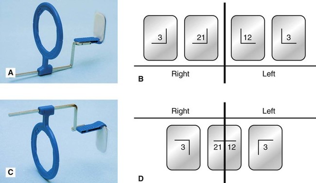
Which exposure techniques require the use of some type of film holder when periapical radiographs are being made. Dental x-ray film processing bydrmeelu lamba pg 1st year oral medicine and radiology 1 2. Cone-cuts appear as a clear zone on traditional radiographs after processing due to the lack of x-ray exposure of the emulsion.

How Make Periapical X Ray

Periapical Radiography Pocket Dentistry

Periapical Radiography Pocket Dentistry

Periapical Radiography Pocket Dentistry

Periapical Radiography Pocket Dentistry

Periapical Radiography Pocket Dentistry
Periapical Radiography Pocket Dentistry
Periapical Radiography Pocket Dentistry
0 comments
Post a Comment